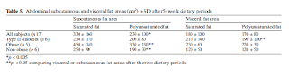
Response of plasma ASP to a prolonged fast
Response of plasma ASP to a prolonged fast - Abstract only unfortunately OBJECTIVE: To determine the changes in the plasma level of acylation stimulating protein (ASP) during a one month total fast in female subjects with marked obesity. DESIGN: Patients with marked obesity underwent a month total fast, before, during (2 weeks), and at the end of which, a variety of relevant metabolic parameters were measured. SETTING: A metabolic unit of a teaching hospital. SUBJECTS: 10 women with marked obesity were studied and the results compared with those in 16 age-matched controls. MAIN OUTCOME MEASURES: Plasma ASP, lipoprotein lipids, apoB, free fatty acid, and ketone levels. RESULTS: At baseline, fasting levels of ASP in the obese group were double that in control subjects (116 +/- 26 vs 53 +/- 30 nM P < 0.001). During the fast, ASP levels dropped progressively and were within the normal range at the end of the study (63 +/- 16 vs 53 +...